Using Qube 384, we profiled a panel of NaV inhibitors across species, providing valuable translational insight early in analgesic drug discovery.
Understanding how drug candidates affect neuronal function within intact neural circuits is a critical step in central nervous system (CNS) drug discovery. Brain slice electrophysiology provides a physiologically relevant platform for investigating compound effects on neuronal excitability, synaptic transmission and network activity in intact brain tissue.
Unlike isolated or cultured cells, brain slices maintain functional neuronal connectivity and local circuitry, enabling a more physiologically relevant assessment of compound effects. This allows researchers to gain valuable insight into ion channel function, synaptic transmission, neuronal firing and overall excitatory activity within a controlled experimental environment.
By bridging the gap between reductionist cell-based assays and in vivo models, brain slice electrophysiology studies play an important role in target validation, mechanism of action investigations and compound optimisation. These studies can increase confidence in candidate progression decisions and improve the likelihood of successful therapeutic outcomes.
Metrion combines extensive ion channel expertise with specialist neuroscience capabilities to deliver high-quality brain slice electrophysiology studies for CNS drug discovery programmes.
Our scientists have extensive experience performing manual patch clamp recordings across multiple brain regions, enabling detailed investigation of neuronal excitability, synaptic transmission and circuit-level pharmacology in physiologically relevant tissue preparations.
Our brain slice assay capabilities provide:
By generating high-quality functional data in intact neural circuits, Metrion helps clients improve confidence in target validation, compound progression decisions and translational neuroscience research.
Electrophysiological studies performed using freshly prepared rodent brain slices, enabling investigation of neuronal and synaptic function within intact neural circuits.
Endpoints can include:
Studies can be tailored to specific targets, mechanisms, disease areas and compound classes to support discovery, translational and mechanistic neuroscience programmes.
Brain slice electrophysiology studies can support:

Figure 1. Hippocampal spontaneous post-synaptic currents (sPSCs) and current-clamp recordings of action potential firing. A. Representative bright field image of a rodent hippocampus, with the subfields cornu ammonis 1 (CA1) and 3 (CA3), and dentate gyrus (DG) indicated. A representative neuron from CA1 (indicated within the box) is shown with a glass recording electrode attached during an experiment (right panel). B. Representative recordings of spontaneous post-synaptic currents at -70 mV holding potential. The inset illustrates a zoomed-in area of the recording. C. Action potential responses to 1-second pulses of injected current as indicated in the current-clamp protocol (upper panel).
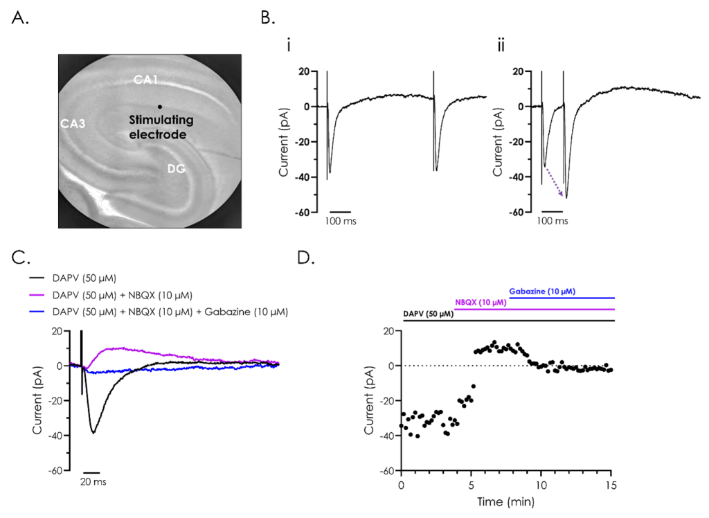
Figure 2. Pharmacological and electrophysiological characterisation of hippocampal evoked post-synaptic currents.
A. Representative bright field image of a rodent hippocampus (left panel), with the subfields cornu ammonis 1 (CA1) and 3 (CA3), and dentate gyrus (DG) indicated. A stimulating electrode was attached close to CA1, as illustrated.
B. A representative recording of evoked post-synaptic currents from hippocampal neurons from CA1, utilising ‘paired-pulse’ stimulation to investigate synaptic plasticity. The effect of reducing the inter-stimulus interval from 500 (i) to 100 (ii) ms illustrates synaptic facilitation, whereby the amplitude of the second evoked current is enhanced as illustrated by the dashed arrow in panel ii.
C. Representative recordings of evoked post-synaptic currents illustrating the different components which make up evoked post-synaptic currents. Recordings were performed after exposure to the NMDA receptor inhibitor D (-)-2-amino-5-phosphonvalerate (DAPV), both alone and in combination with the AMPA receptor inhibitor 2,3-dioxo-6-nitro-7-sulfamoyl-benzo(f)quinoxaline (NBQX), and GABA antagonist, gabazine.
D. A representative current-time plot showing the peak inward and outward currents recorded from the different components of the evoked post-synaptic currents during a 15-minute recording.
Brain slice tissue assays use thin sections of freshly prepared brain tissue to investigate neuronal and synaptic function while preserving native brain architecture and connectivity.
Brain slices retain neuronal networks, synaptic connections and local circuit organisation that are often lost in dissociated cell culture systems, providing a more physiologically relevant model for studying CNS pharmacology and neuronal function.
Metrion primarily uses gold-standard manual patch clamp electrophysiology to investigate neuronal excitability, ion channel activity, synaptic transmission and pharmacological responses within brain slice preparations.
Endpoints include intrinsic firing properties, action potential generation, spontaneous and evoked synaptic responses, neuronal excitability, synaptic transmission and synaptic plasticity.
Brain slice electrophysiology studies can support programmes focused on epilepsy, pain, neurodegenerative diseases, neuropsychiatric disorders and other CNS indications where neuronal and synaptic function are important drivers of disease biology.
Brain slice assays provide mechanistic insight into compound effects within intact neural circuits, helping researchers understand target biology, evaluate pharmacological activity and make more informed compound progression decisions.
Using Qube 384, we profiled a panel of NaV inhibitors across species, providing valuable translational insight early in analgesic drug discovery.
We explore hNav1.9's unique fast and slow inactivation properties using Qube 384 and QPatch 48 platforms, helping to build more predictive screening assays for state-dependent inhibitors.