Expanding our high-throughput ion channel screening capabilities.
Metrion provides hiPSC-derived cardiomyocyte screening services to help drug discovery and safety pharmacology teams identify potential proarrhythmic liabilities early in development.
Using human induced pluripotent stem cell (hiPSC)-derived cardiomyocytes and gold-standard manual patch-clamp electrophysiology, our scientists assess the effects of compounds on cardiac action potentials and key electrophysiological parameters. These studies support the evaluation of proarrhythmic risk, helping clients make informed progression decisions before costly later-stage development.
Our cardiac safety experts generate high-quality electrophysiology data that can distinguish compounds with low, medium, or high proarrhythmic risk profiles. The approach aligns with industry best practice and supports broader cardiac safety assessment strategies, including investigations related to CiPA (Comprehensive in vitro Proarrhythmia Assay) initiatives.
In drug discovery, identifying potential heart-related side effects early in the process is crucial to ensuring the safety of new therapeutic compounds. Evaluation of the tendency of a compound to cause abnormal heart rhythms, which can lead to life-threatening arrhythmias is a key assessment. Accurately identifying compounds with low, medium, or high proarrhythmic risk profiles is valuable in reducing the likelihood of cardiovascular complications in patients. An important method for assessing the proarrhythmic potential of drug candidates that has been recognised for its value by the Regulatory authorities is the evaluation compound effects on action potentials (APs) morphology in cardiomyocytes.
Human-induced pluripotent stem cell (hiPSC)-derived cardiomyocytes offer a cutting-edge, human-relevant model for this kind of testing. By recording spontaneous action potentials from these cells, we can monitor the impact of compounds on key parameters such as action potential duration (APD), amplitude, and morphology. With this data, we can detect subtle changes that may indicate potential arrhythmic risks. Using the gold-standard manual patch-clamp technique we can obtain high-fidelity results that help distinguish between compounds with varying proarrhythmic risk profiles. This approach not only offers valuable insights into drug safety but also improves understanding of the mechanisms behind drug-induced arrhythmias.
Metrion combines deep expertise in cardiac electrophysiology, ion channel pharmacology and safety pharmacology to deliver high-quality hiPSC-derived cardiomyocyte screening studies. Our team works closely with clients to generate reliable, decision-enabling data that supports compound progression and cardiac safety risk assessment.
One of the most powerful techniques for assessing the proarrhythmic potential of drugs is the manual patch-clamp method. This gold-standard electrophysiological technique allows for precise measurement of the electrical activity of individual cells, making it possible to detect even subtle changes in the action potential waveform that may indicate potential arrhythmogenic effects. In the current clamp format, we can record action potentials from a single cardiomyocyte, whereby Metrion scientists can record the cell’s response in real-time, capturing the characteristic shape and duration of the action potential.
The action potential parameters recorded during the assay include:
By assessing these parameters, Metrion scientists can evaluate how drugs affect the cell’s electrophysiological properties. The manual patch-clamp technique is particularly effective at detecting subtle changes to these parameters, making it ideal for identifying compounds with varying levels of proarrhythmic risk.
One of the major advantages of using the manual patch-clamp assay with hiPSC-derived cardiomyocytes is its ability to provide continuous, real-time monitoring of drug effects over time. The recordings are stable for more than 30 minutes, allowing researchers to observe the cumulative effects of multiple concentrations of a compound.
This time window enables the examination of how the compound behaves over a prolonged exposure period, which is particularly important for drugs that may cause delayed or cumulative effects. By applying various concentrations of the drug during the assay, researchers can determine the dose-dependent effects on action potential parameters and discern compounds that pose a higher or lower risk for proarrhythmia.
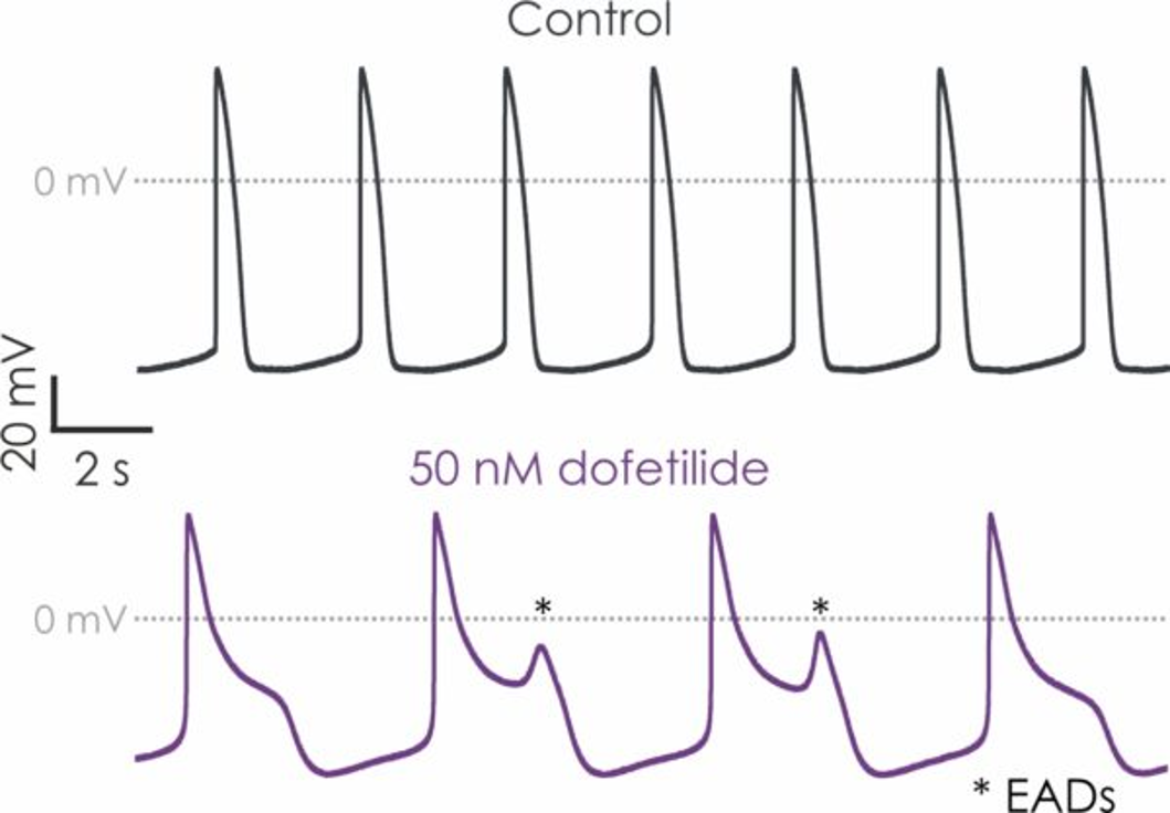
To illustrate the utility of this assay, let’s consider the example of dofetilide, a drug used to treat atrial fibrillation. Dofetilide is known to prolong APD, which increases the risk of arrhythmias. Using the manual patch-clamp assay with hiPSC-derived cardiomyocytes, we can measure the impact of dofetilide on APD and other action potential parameters.
When exposed to 50 nM of dofetilide, the compound provoked early afterdepolarisations (EADs) and caused a significant prolongation of all measured APD values in hiPSC-derived cardiomyocytes (Figures 1 and 2). This is a clear indicator of the proarrhythmic potential of dofetilide. In addition, the stability of the spontaneous beats from a cardiomyocyte becomes disturbed after exposure to dofetilide, in comparison with vehicle-treated controls (Figure 3).

Figure 1. hiPSC-derived cardiomyocyte action potential screening.

Figure 2. Drug effects on hiPSC-derived cardiomyocyte responses.

Figure 3. Changes in hiPSC-derived cardiomyocyte beat stability.
Proarrhythmia is a serious concern in drug development, as many pharmaceuticals - particularly those targeting the heart or central nervous system - can alter the normal rhythm of the heart. These drugs may affect the electrical properties of cardiomyocytes, leading to prolonged or shortened action potentials, delayed repolarisation, or other disturbances that can result in arrhythmias.
Identifying drugs that pose a proarrhythmic risk early in the development process is critical to reducing harm and improving patient safety. Traditional in vivo models are often inadequate in predicting human-specific cardiac responses. They fail to capture the complexity of human cardiac electrophysiology and often lead to unreliable or inconclusive results. This is where hiPSC-derived cardiomyocytes are can play a key role for testing proarrhythmic potential.
hiPSC-derived cardiomyocytes are stem cell-derived heart cells that mimic human cardiac function more closely than animal-derived cells. These cells enable researchers to examine drug effects on human-specific cardiac ion channels and electrophysiological behaviour, giving them more accurate and reliable data than conventional models. By evaluating drug effects on these human-relevant cells, researchers can better predict the likelihood of arrhythmias and reduce the risk of adverse cardiovascular events.
hiPSC-derived cardiomyocyte assays are increasingly used as part of modern cardiac safety assessment strategies and can provide valuable supporting data for Comprehensive in vitro Proarrhythmia Assay (CiPA)-related investigations. Read more about CiPA screening.
hiPSC-derived cardiomyocyte screening is a cardiac safety assay that uses human stem cell-derived heart cells to evaluate how drug candidates affect cardiac electrophysiology and proarrhythmic risk.
Key endpoints include action potential duration (APD), action potential amplitude, upstroke velocity, repolarisation characteristics, beat stability, and the occurrence of early afterdepolarisations (EADs).
Manual patch clamp remains the gold standard for action potential recording, delivering high-quality electrophysiological data and detailed mechanistic insight into compound effects on cardiac function.
These studies help identify potential cardiac safety liabilities earlier in development, supporting candidate selection, risk assessment, and regulatory decision-making.

Expanding our high-throughput ion channel screening capabilities.
Biologic modalities may offer opportunities to achieve levels of subtype selectivity that can be difficult to obtain using traditional small molecules, particularly across closely related ion channel family members. This may enable more precise modulation of disease-relevant targets while potentially reducing unwanted off-target activity.