Action Potential Waveform Analysis in Human IPSC-Cardiomyocytes Enables Mechanistic Assessment of Multichannel Cardiac Effects

Graham D. Smith, Katie L. Puddefoot, Sergiy Tokar, Alexander Haworth, Catherine M. Hodgson, Thomas D. M. Hill, Ayesha Jinat, Anthony M. Rush, Edward B. Stevens, Gary S. Clark
Metrion Biosciences Ltd, Building 2, Granta Centre, Granta Park, Cambridge, CB21 6AL, UK
Encoded by the SCN11A gene, NaV1.9 is a voltage-gated sodium (NaV) channel highly expressed in trigeminal ganglion neurons and small-diameter nociceptors in the dorsal root ganglion. NaV1.9 acts as a threshold channel with a lower activation threshold, slower biophysical properties and a large window current compared to the other NaV isoforms1. These characteristics are important for its role in the regulation of neuronal excitability and the modulation of inflammatory and neuropathic pain. Clinically, NaV1.9 dysfunction has been implicated in altered pain perception in humans (Figure a) evidencing its potential as a non-opioid pain target2,3,4.
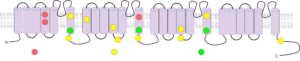
Figure a – Schematic diagram of pain-related mutations in hNaV1.9, adapted from Kabata et al.5. Red: Familial episodic pain. Yellow: Painful peripheral neuropathy. Green: Congenital insensitivity to pain.
High-throughput NaV1.9 drug discovery programmes have been hindered to date by the lack of the cellular tools currently available. Hence, the generation of a robust, stably-expressing hNaV1.9 cell line and high-throughput assay would greatly accelerate the development of selective NaV1.9 modulators without the side-effects associated with current pain treatment options.
A stably-expressing monoclonal CHO-hNaV1.9 cell line was generated in-house at Metrion Biosciences.
CHO-hNaV1.9 cells were stimulated from ‑100 mV to +50 mV (50 ms, 10 mV steps) from a holding potential of ‑140 mV, at 0.05 Hz.
For IV analysis, CHO-hNaV1.9 cells were stimulated similar to the manual patch protocol, except 100 ms per step. For compound screening, cells were stimulated with a single 50 ms step to ‑40mV from ‑140 mV (0.05 Hz). Automated recordings were performed using multi-hole on a Qube 384 (Sophion Bioscience) in the presence of 300 nM TT.

Figure 1 - Monoclonal CHO-hNaV1.9 cell lines were generated using dilution cloning and screened using a FLIPR Penta system. Positive clones were subsequently screened using a Qube 384 automated patch clamp platform, with the best further validated using manual patch clamp.
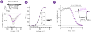
Figure 2 - Representative current traces and IV analysis of hNaV1.9, recorded using the whole-cell manual patch clamp technique (A). Conductance/voltage plot for hNaV1.9 (B). hNaV1.9 currents were inhibited using the NaV channel blocker, TC-N 1752, at 3 µM (C). The selected monoclone was confirmed to express hNaV1.9 and was progressed to assay development on the Qube automated electrophysiology platform.
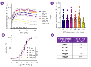
Figure 3 - Enhanced G-protein signalling has been shown to potentiate NaV1.9 current amplitudes6. Addition of up to 500 µM intracellular GTPγS resulted in larger hNaV1.9 currents, whilst 2000 µM GTPγS reduced hNaV1.9 current amplitude (A,B). Importantly, GTPγS concentration did not alter hNaV1.9 pharmacology (C,D). A concentration of 200 µM was selected for future experiments.
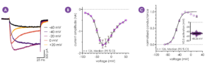
Figure 4 - NaV1.9 channels have distinct biophysical properties compared to the other NaV isoforms1 (A). The IV relationship (B) and conductance (C) of hNaV1.9 currents recorded from 126 Qube 384 multi-hole wells were consistent with the known characteristics of native hNaV1.9 and data obtained using the manual patch clamp technique.
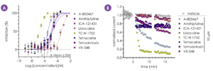
Figure 5 - A Qube 384 assay was successfully developed to screen a selection of NaV inhibitors with a range of potencies and isoform selectivity against hNaV1.9. Calculated IC50 values (µM): A-803467 – 3.51, Amitriptyline – 18, ICA-121431 >30, Lidocaine – 460, TC-N 1752 – 0.2, Tetracaine – 11.5, TTX – 18.7, VX-548 – 14.1 (A). Representative I-t plots are shown in B.
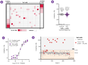
Figure 6 - The robustness of the Qube 384 assay was further validated by assessing the potency of TC-N 1752, using a randomised spiked plate approach (plate map - A). Vehicle response and TC-N 1752 potency correlated well between control and test wells (B, C). In test wells, the vehicle response displayed low variability with the TC-N 1752 response (at >0.1 µM) easily discernible above mean vehicle response + 3 SD threshold (D).
Action Potential Waveform Analysis in Human IPSC-Cardiomyocytes Enables Mechanistic Assessment of Multichannel Cardiac Effects
Cardiac toxicity remains the leading cause of new drug safety side-effects. Current preclinical cardiac safety assays rely on in vitro cell-based ion channel assays and ex vivo and in vivo animal models⁽¹⁾. These assays provide an indication of acute risk but they do not always predict the effect of chronic compound exposure, as recently seen with oncology drugs. Therefore, new assays are required to characterise chronic structural and functional effects in human cells earlier in drug discovery. Impedance-based technology can provide more accurate chronic cardiotoxicity measurements in an efficient manner using human induced pluripotent stem cell-derived cardiomyocytes (hiPSC-CMs).