A stable cell line expressing KV3.1 V434L variant was developed and characterised, confirming published data describing V434L as a gain-of-function mutation.
Robert W. Kirby, Said El Haou, Edward Humphries, John Ridley and Marc Rogers
Cardiac toxicity remains the leading cause of new drug safety side-effects. Current preclinical cardiac safety assays rely on in vitro cell-based ion channel assays and ex vivo and in vivo animal models⁽¹⁾. These assays provide an indication of acute risk but they do not always predict the effect of chronic compound exposure, as recently seen with oncology drugs⁽²⁾. Therefore, new assays are required to characterise chronic structural and functional effects in human cells earlier in drug discovery⁽²̛ ³⁾. Impedance-based technology can provide more accurate chronic cardiotoxicity measurements in an efficient manner using human induced pluripotent stem cell-derived cardiomyocytes (hiPSC-CMs)⁽¹̛ ³⁾.
Here we outline validation of chronic cardiotoxicity assays using an impedance platform in combination with commercial hiPSC-CMs:
hiPSC-CMs from commercial vendors (Ncardia and CDI) were plated onto CardioExcyte96 well plates (Nanion) according to manufacturers’ instructions. Plates were incubated at 37 °C (5 % CO2 ) for 7-10 days and 100 % medium exchanges were performed daily. Impedance signals were recorded using the CardioExcyte96 (CE96) platform and the formation of a spontaneous beating syncytial network was monitored with 30 s impedance recording every hour (1 hr) during the first week in culture.
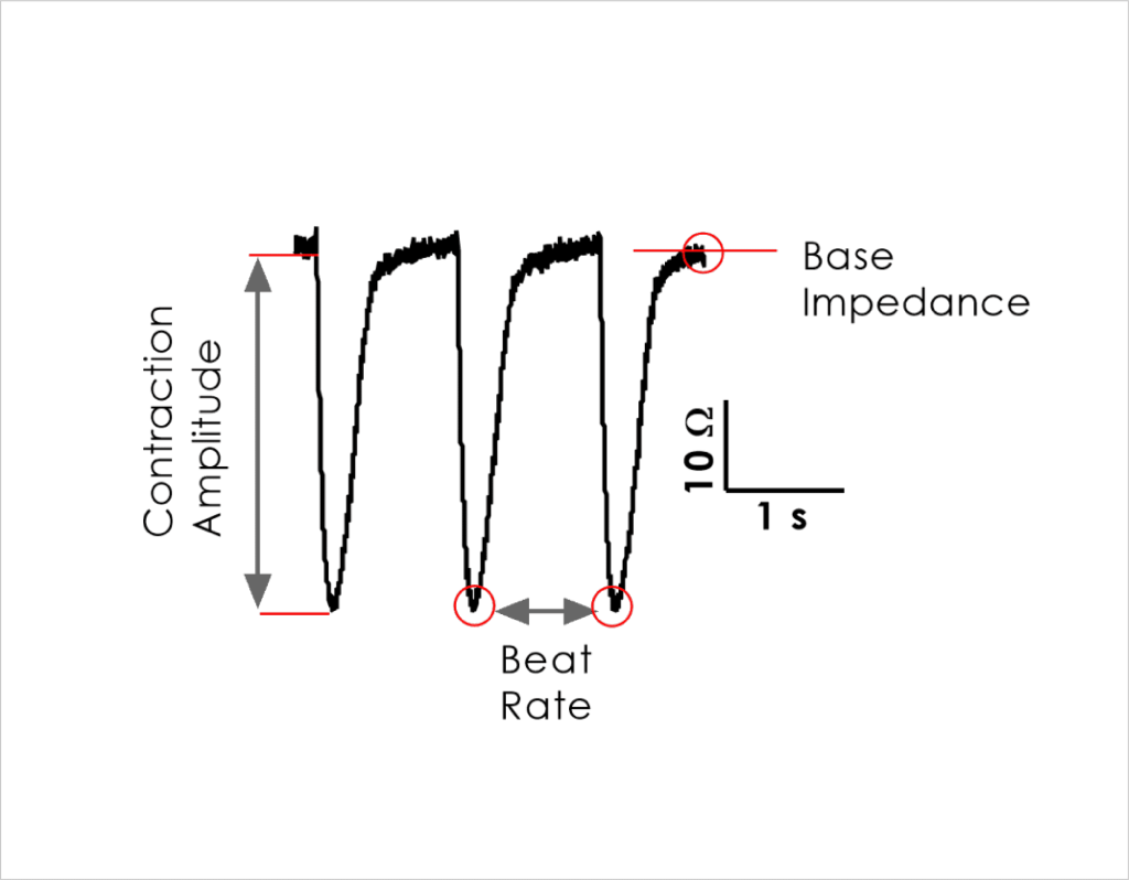
A single concentration of compound was then applied to each well (N ≥ 4) every 24 hrs as a 1:1000 fold dilution into media (final = 0.1 %) for a maximum of 72 hours before a washout period. The outputs (Figure 1) were recorded at 60 minute intervals to assess acute vs. chronic effects (< 24 hrs vs 24 – 72 hrs).
Impedance assay
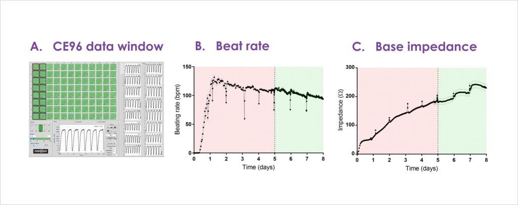
hiPSC-CMs from a commercial vendor plated onto a CardioExyte96 well plate delivered a high success rate across the plate
(A) Greater than 90% of wells started beating within 6 hours and stabilised within 72 hours
(B) Consistent base impedance (growth phase) parameters were obtained after 5 days
(C) Recordings were suitable for chronic drug screening after 4-7 days
Cardiac ion channel modulator effects on contractility
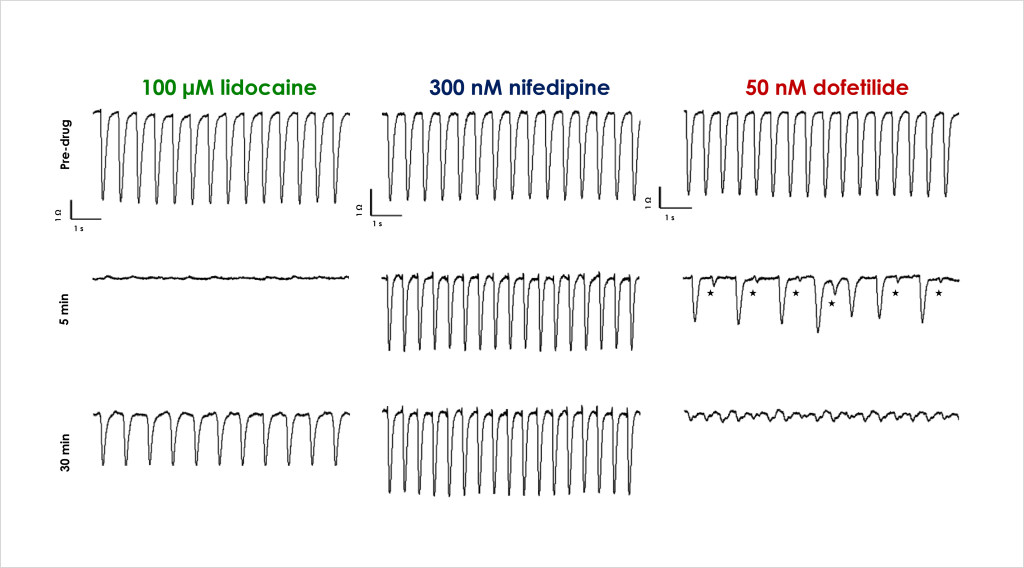
A. Acute effects of ion channel ligands on cell contraction
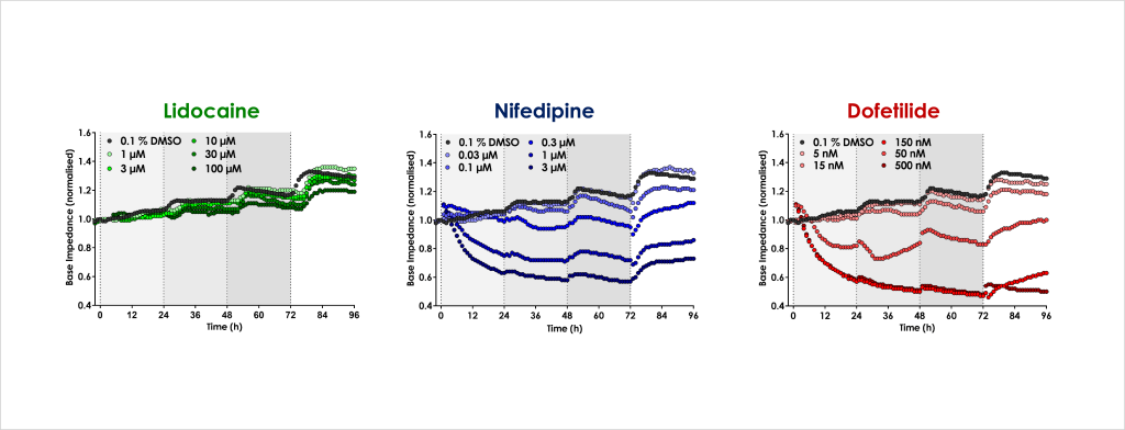
B. Chronic cardiotoxicity drug effects
(A): Cardiac contractility was assessed by examining changes in beat rate and amplitude. The expected acute effects of cardiac ion channel modulators of IKr (dofetilide, 50 nM), INa (lidocaine, 100 µM) and ICa (nifedipine, 300 nM) were confirmed, as represented by the traces above showing spontaneous contractile activity in control conditions (Pre-drug) and in presence of drug after 5 and 30 min incubation. Lidocaine stopped contraction but beating recovered with a decreased beat rate & amplitude, nifedipine increased beat rate but decreased amplitude, and dofetilide decreased amplitude & beat rate with secondary beats degenerating into fibrillation.
(B): Chronic cardiac drug effects on base impedance Base impedance was used to monitor cell viability over 72 hour period. Lidocaine had no effect at 72 hrs relative to control whereas nifedipine and dofetilide produced time- and concentration-dependent decreases in base impedance within 24 hrs.
Chronic cardiotoxicity mechanisms
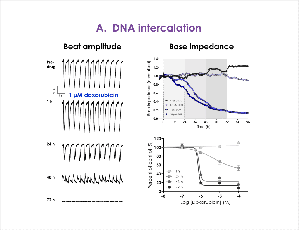
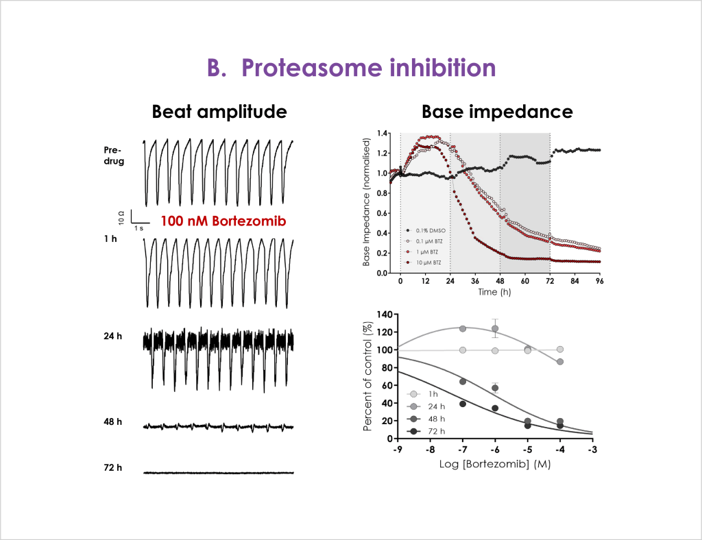
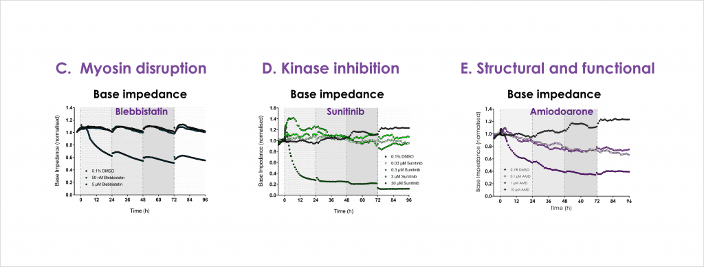
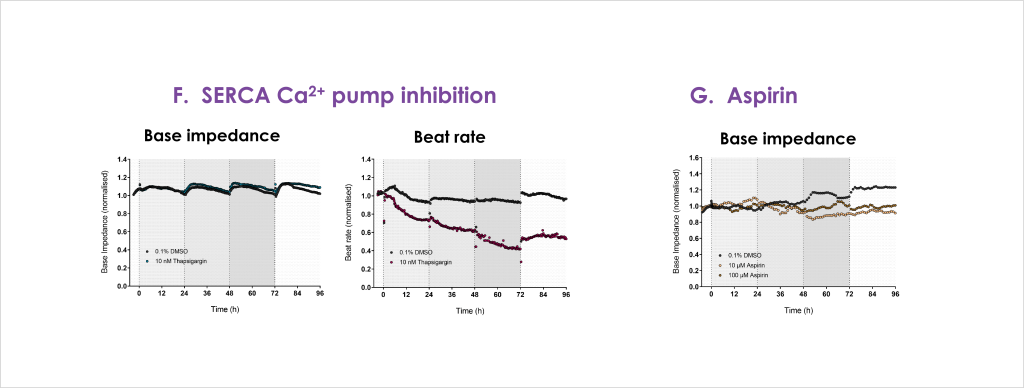
Functional and structural cardiotoxic effect of drugs with different modes of action, including several therapeutic oncology reagents, were tested. Drug mechanisms included DNA intercalation (doxorubicin), proteasome inhibition (bortezomib), myosin disruption (blebbistatin), SERCA Ca2+ pump inhibition (thapsigargin), kinase inhibition (Sunitinib), and an ion channel modulator with mixed functional and structural effects (amiodarone). Aspirin is considered non-cardiotoxic and was used as a negative control⁽⁴⁾.
A. DNA intercalator
B. Proteosome inhibitor
C. Myosin disruptor, D. Kinase inhibitor and E. Structural and functional inhibitor
F. SERCA Ca2+ pump inhibitor and G. Negative control
Concentration and time-dependent decreases in base impedance were observed for doxorubicin with > 24 hrs required to resolve the full effect (A). Bortezomib produced biphasic effects, initially increasing base impedance (< 24 hrs) at all concentrations tested, before impairment of contractility was seen at > 24 hrs (B). The myosin disruptor blebbistatin produced a rapid and sustained reduction in base impedance within 24 hrs (C). Similar to bortezomib, the oncology kinase inhibitor sunitinib produced a transient and dose-dependent increase in base impedance at 3 and 30 µM before longer incubation and higher concentrations disrupted base impedance (D). The non-specific ion channel modulator amiodarone also produced concentration and time-dependent deficits in cell impedance (E). The Ca2+ pump inhibitor thapsigargin showed a small tendency to increase base impedance, and as expected more pronounced effects on beat rate contractility due to depletion of intracellular calcium stores (F). The negative control Aspirin showed little concentration or time-dependent effects on cell impedance (G).
H. Atypical cardiac toxicity of Trastuzumab
Trastuzumab is a monoclonal antibody approved for treating HER2-positive breast and gastric cancer. However, clinical incidents and post-market analysis reveal that therapeutic use has been limited by the occurrence of adverse cardiac events, the mechanism(s) of which are not fully realised⁽⁵⁾.
Figure 5: Trastuzumab (0.5 – 50 µg/ml) was applied at 0, 24, 48 and 72 hrs after base impedance stability was achieved. Significant effects on base impedance relative to control were only recorded > 72 hrs following multiple applications of antibody, suggesting either that entry into the cell is limited or the modulation of substrates responsible for this cardiotoxicity is slowacting.
A stable cell line expressing KV3.1 V434L variant was developed and characterised, confirming published data describing V434L as a gain-of-function mutation.
The development and validation of electrophysiological assays to study TRPML1 is important to understand the function and pharmacology of the channel. We used a TRPML1 variant that lacks the endo-lysosomal retention sequences (TRPML1-4A), enabling the channel to express at the plasma membrane3. As such channel behaviour can be characterised by means of whole-cell patch-clamp and fluorescence-based techniques.