Development and Evaluation of Novel Solution Pairs to Enhance Seal Resistance in Automated Patch Clamp Electrophysiology and Ion Channel Experiments

Authors

Catherine Hodgson1, Edward Stevens1, Andrew James1, Anissa Bara2, Beatrice Badone2, Damian Bell2, Daniel Sauter2, Gary Clark1

1Metrion Biosciences Ltd, Granta Centre, Granta Park, Cambridge, CB21 6AL, UK

2Sophion Bioscience A/S, Industriparken 39, 2750 Ballerup, Denmark

Introduction

Overview of automated patch clamp electrophysiology and ion channels

Gigaohm seals, or ‘gigaseals’, are crucial for patch clamp electrophysiology, ensuring excellent electrical access to cells to enable high-quality recordings. These seals form through chemical bonds and electrostatic forces between the cell membrane and the glass pipette in manual patch clamp, or between the cell membrane and chip substrate in planar patch clamp. Planar patch clamp often employs ‘seal enhancers’ to increase seal resistances, with CaF2 being the most commonly used. It is hypothesized that high extracellular Ca2+ and intracellular F- concentrations lead to CaF2 precipitate formation at the solution interface, promoting seal formation.

However, CaF2 as a seal enhancer has limitations. F- interacts with various internal components such as protein kinase A, adenylate cyclase, and lipid phosphatases, which can affect the biophysical properties of some ion channels. Additionally, F- is not ideal when recording from Ca2+- activated ion channels due to the resulting unknown concentrations of free intracellular Ca2+.

In an effort to overcome these limitations, Sophion developed new solution pairs in 2017 that foster seal formation (Patent: WO2018100206A1). Building on this technology, Metrion and Sophion collaborated to further determine whether other insoluble salts can act as seal enhancers and how these solution pairs affect the biophysical properties and pharmacology of the investigated ion channels.

Materials and Methods

Materials and methods for gigaseal patch clamp electrophysiology experiments

Experiments were conducted using a Sophion Bioscience Qube 384 with QChip 384 (single hole) and QChip 384X (multihole) consumables. Temperature was maintained at 22 °C using the Qube temperature control module.

Analysis was conducted using Sophion Analyzer v9.0.42 and GraphPad Prism v10.2.2.

CHO-hNav1.5 and HEK293-hCav1.2 cell lines were provided by Metrion Biosciences.

All compounds were tested at: 0.001, 0.01, 0.1, 1, 10 and 100 µM.

Results

Results from automated patch clamp electrophysiology and gigaseal studies

Table 1. Correlation between salt pair solubility product constants (Ksp) and gigaseal formation on Qube 384. Little correlation was found between the solubility product constants (Ksp) of Ca2+, Ba2+ and Sr2+ salts (A) and their ability to foster gigaseal formation (B). Despite the PO­43- salts having very low Ksp values and SrCO3 having a similar Ksp value to CaF­2 and BaSO4, these salts failed to produce gigaohm seals. Moderate seal resistances with PO43- salts were transient and unstable. Median resistances calculated from 24 cells per salt pair.

Correlation between salt pair solubility product constants (Ksp) and gigaseal formation on Qube 384

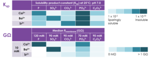
Table 2. Resistances of seals formed using various concentrations of extracellular Ba2+ and intracellular SO42-. Gigaseals only formed with ≥ 3 mM Ba2+ and in the presence of SO42-. Median resistances calculated from 24 or 48 cells per condition.

Resistances of seals formed using various concentrations of extracellular Ba2+ and intracellular SO42

Effects of CaF2 and BaSO4 on hNav1.5 channel biophysics

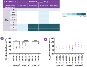
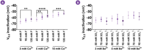
Figure 1. Effects of CaF2 and BaSO4 on hNav1.5 channel biophysics. hNav1.5 V0.5 inactivation with different cation and anion concentrations (mean ± S.D.; N ≥ 11). Increasing concentrations of intracellular F- caused a depolarising shift in V0.5 inactivation (A). In contrast, increasing concentrations of SO42- had no effect on hNav1.5 V0.5 inactivation (B). One-way ANOVAs conducted within each cation group followed by Tukey’s Honestly Significant Difference post-hoc tests: ** = p < .01; *** = p < .001; **** = p < .0001.

CaF2 versus BaSO4 – hNav1.5 pharmacology

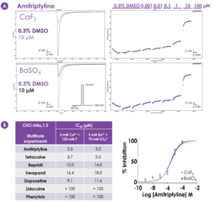
Figure 2. CaF2 versus BaSO4 – hNav1.5 pharmacology. A) Representative sweep plots (left) and current-time (I-t) plots (right) for hNav1.5 inhibition by amitriptyline. There was no difference in cumulative inhibition of hNav1.5 by increasing concentrations of amitriptyline between CaF2 and BaSO4. B) Screening of a range of inhibitory compounds showed no difference in hNav1.5 pharmacology between CaF2 and BaSO4. Concentration-response curves for amitriptyline against hNav1.5 using CaF2 or BaSO4 as the seal enhancer (mean ± S.D.; N = 12 wells per concentration for CaF2, N = 8 wells per concentration for BaSO4).

CaF2 versus BaSO4 – hCav1.2 kinetics

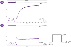
Figure 3. CaF2 versus BaSO4 – hCav1.2 kinetics. hCav1.2 exhibits Ca2+-dependent inactivation when CaF2 is used as the seal enhancer (A). BaSO4 as the seal enhancer (using Ba2+ as a surrogate carrier ion) (B) confers loss of the Ca2+-dependent inactivation of hCav1.2 observed with CaF2. Example sweep plots derived from Sophion Analyzer v9.0.42.

CaF2 versus BaSO4 – hCav1.2 pharmacology

Figure 4. CaF2 versus BaSO4 – hCav1.2 pharmacology. Concentration-response curves for two common inhibitors against hCav1.2, nifedipine (A) and verapamil (B) (CaF2: N = 2‑5 wells per concentration; BaSO4: N = 6-12 wells per concentration). Compound potencies (IC50 values) did not differ between CaF2 and BaSO4. Data displayed as mean ± S.D.


Conclusions

Findings of automated patch clamp electrophysiology experiments

  • BaSO4 was identified as an equivalent seal enhancer to CaF2
  • BaSO4 and CaF2 characterized across two ion channels: hNav1.5 and hCav1.2.
  • Intracellular F- caused depolarising shifts in the voltage dependence of inactivation of hNav1.5 where no such effects were observed with SO42- in the intracellular solution.
  • No difference in pharmacological effects of inhibitory compounds against hNav1.5 or hCav1.2 was observed between the two solution pairs, CaF2 and BaSO4.
  • BaSO4 is well-suited as a seal enhancer for recording from non-K+-conducting Ca2+-activated channels, such as TMEM16A. In particular as BaSO4 allows more accurate estimation of free intracellular Ca2+ concentration.
Recommended Publications
Latest Publications
Development of a High-Throughput Automated Electrophysiology Assay for Human NaV1.9 Inhibitor Screening in Neuropathic Pain Research

A stable cell line expressing KV3.1 V434L variant was developed and characterised, confirming published data describing V434L as a gain-of-function mutation.

High-throughput Drug Repurposing Screen of the Potassium Channel KV3.1, with V434L Mutation (case study)

A stable cell line expressing KV3.1 V434L variant was developed and characterised, confirming published data describing V434L as a gain-of-function mutation.

View All
Metrion Biosciences is a contract research organisation (CRO) specialising in high-quality preclinical drug discovery services.
magnifier
linkedin facebook pinterest youtube rss twitter instagram facebook-blank rss-blank linkedin-blank pinterest youtube twitter instagram