Action Potential Waveform Analysis in Human IPSC-Cardiomyocytes Enables Mechanistic Assessment of Multichannel Cardiac Effects

Authors

Charlotte Hill and Robert Kirby

  • Metrion Biosciences Ltd., Cambridge, UK

Introduction

Recent regulatory changes, including the FDA Modernisation Act 2.0 and the UK government’s Replacing Animals in Science strategy, have accelerated the adoption of human-relevant New Approach Methodologies (NAMs) or Non-Animal Alternatives (NAAs) for nonclinical safety assessment. In cardiac safety evaluation, reliance on single ion channel assays, such as the rapid delayed rectifier potassium channel hERG (KV11.1), may fail to capture the integrated electrophysiological effects that underlie proarrhythmic risk.

Parameters derived from action potential waveform analysis, including triangulation, engagement of repolarisation reserve and presence of early afterdepolarisations (EADs), are recognised as contributors to arrhythmogenic liability. Human induced pluripotent stem cell derived cardiomyocytes (hiPSC-CMs) provide a translationally relevant system in which integrated multichannel cardiac effects can be assessed.

Aim

The aim of this study was to evaluate whether optical voltage imaging of hiPSC-CM action potential waveforms can detect compound-induced electrophysiological changes beyond hERG block alone and enable mechanistic interpretation through correlation with ion channel electrophysiology data.

Materials and Methods

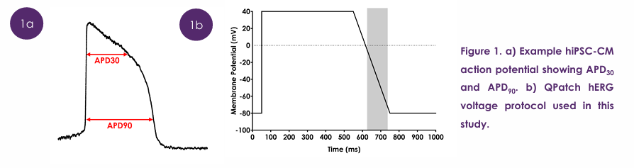
hiPSC-CMs (iCell2; Fujifilm CDI) were seeded into 96-well plates. After 8 days, cells were loaded with voltage-sensitive dye (BeRST1) and placed in the Lumencor VOLTA scanner, set to 28°C. Action potentials were measured optically for 40 s using fluorescence (10 kHz; excitation 660 nm/emission 680 nm). Compounds were applied (10 concs; 0.02 nM – 30 µM), incubated for 30 min and 24 h, followed by a 40 s recording. Clinical QTc risk analysis of ycAPD903 was performed using the thresholds and equations according to Kilfoil et al., 20212. Triangulation was calculated per well as APD30/APD90 and normalised to vehicle (0.1% DMSO), where a decrease in ratio indicates an increase in triangulation (Figure 1 a). Whole cell voltage clamp experiments were performed at 23°C on CHO cells stably expressing hERG using the Sophion QPatch48 platform. Recording solutions: extracellular (in mM) NaCl 140, KCl 2, CaCl2 2, MgCl2 1, HEPES 10, glucose 5; intracellular (in mM) KF 120, KCl 20, HEPES 10, EGTA 10. Currents were elicited using the voltage protocol shown in Figure 1 b.
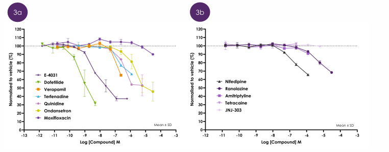
  • The selective hERG inhibitors, dofetilide and E-4031, produced an early and pronounced increase in triangulation (Figure 3 a).
  • Multi-ion channel effect compounds, such as verapamil, terfenadine, quinidine and ondansetron, all showed concentration-dependent increases in triangulation at higher exposures (Figure 3 a), consistent with modulation of inward (e.g. ICaL) and outward (e.g. IKr) currents.

Effect of compounds on triangulation ratio (APD30/APD90)
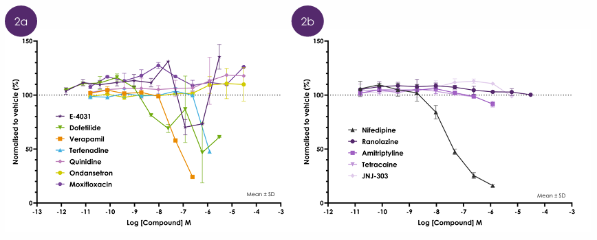
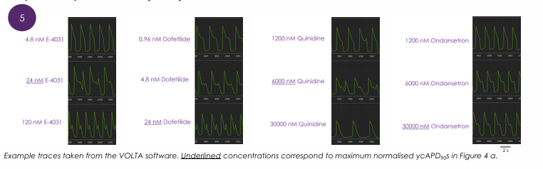
  • Compounds with multi-ion channel effects, such as quinidine and ondansetron showed less pronounced effects on APD30 (Figure 2 a), despite the presence of EADs (Figure 5).

Early afterdepolarisations (EADs)
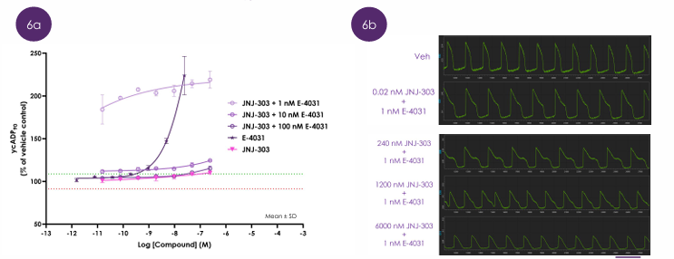
  • Low concentration of E-4031 resulted in reduction in repolarisation reserve by synergistic inhibition of hERG and hKVLQT1/mink to increase APD (Figure 6).
  • Higher concentrations of E-4031 resulted in prolongation of the action potential early in phase 2, which can shift voltage-dependent inactivation kinetics of ICaL and counterbalance further APD prolongation (Figure 6).

Repolarisation reserve and ycAPD903
Results

  • Compound-mediated effects on APD30 were more consistent and more pronounced for some compounds, such as verapamil and nifedipine, indicating reduced inward calcium current (Figure 2).

Effect of compounds on APD30

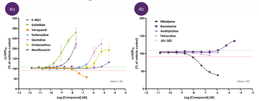
  • Dofetilide and E-4031 increased APD90 and triangulation at low nM concentrations (Figures 3 a and 4 a) but also affected APD30 at µM concentrations (Figure 2 a), which is where EADs were observed in raw traces (Figure 5), indicating instability of the action potential.

Effect of compounds on ycAPD903 after 30 min incubation

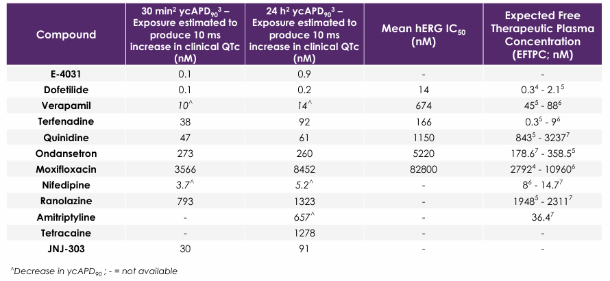
Table 1. Summary of estimated clinical QTc risk, hERG inhibition and EFTPC


Conclusions

  • Action potential waveform analysis in hiPSC-CMs enables mechanistic differentiation of multi-ion channel cardiac effects beyond hERG inhibition and action potential duration, supporting its application in human-relevant cardiac safety assessment.
  • These findings demonstrate that optical voltage imaging of hiPSC-CM action potentials provides mechanistic insight into multi-ion channel cardiac effects that cannot be captured by hERG alone.

References

  1. Huang, Y.L., Walker, A.S., & Miller, E.W. (2015). PMID: 26237573.
  2. Kilfoil, P. et al., (2021). PMID: 34678241.
  3. Yamamoto, W. et al., (2016). PMID: 27923051.
  4. ICH E14/S7B Q&As Training Material Examples Supplemental File (2022).
  5. Crumb, W.J. et al., (2022). PMID: 27060526.
  6. Kramer J. et al., (2013). PMID: 23812503.
  7. Heitmann, S., Vandenberg, J.I., Hill, A.P. (2023). PMID: 38079357
Recommended Publications
Latest Publications
Validation of antibody toxin fusions against sodium channels

Cardiac toxicity remains the leading cause of new drug safety side-effects. Current preclinical cardiac safety assays rely on in vitro cell-based ion channel assays and ex vivo and in vivo animal models⁽¹⁾. These assays provide an indication of acute risk but they do not always predict the effect of chronic compound exposure, as recently seen with oncology drugs. Therefore, new assays are required to characterise chronic structural and functional effects in human cells earlier in drug discovery. Impedance-based technology can provide more accurate chronic cardiotoxicity measurements in an efficient manner using human induced pluripotent stem cell-derived cardiomyocytes (hiPSC-CMs).

Evaluation of hNav1.9 Screening Cascade for Analgesic Drug Discovery

Automated patch clamp enables robust NaV1.9 screening, accelerating discovery of selective sodium channel modulators for pain research.

View All
magnifier
linkedin facebook pinterest youtube rss twitter instagram facebook-blank rss-blank linkedin-blank pinterest youtube twitter instagram