Action Potential Waveform Analysis in Human IPSC-Cardiomyocytes Enables Mechanistic Assessment of Multichannel Cardiac Effects

Catherine M. Hodgson, Scott A. Maidment, Remigijus Lape, Gary S. Clark, Robert W. Kirby, Anthony M. Rush, Edward B. Stevens
KV7.2/7.3 (KCNQ2/3) potassium channels underlie the neuronal M-current, a key regulator of membrane excitability and action potential firing.1 Pharmacological activation of these channels represents a validated strategy for dampening hyperexcitability in disorders such as epilepsy and neuropathic pain.2 Activators of KV7.2/7.3 exhibit differing mechanisms of action, including pore stabilisation and voltage sensor modulation dependent upon lipid cofactors such as phosphatidylinositol 4,5-bisphosphate (PIP2).3,4
We have developed an integrated assay platform combining high-throughput screening using a fluorescent thallium dye (FLIPR® Penta), automated voltage clamp (Qube 384) and current clamp (QPatch 48) electrophysiology, and manual patch clamp recordings from sensory neurons to characterise KV7.2/7.3 activators.
Retigabine5, flupirtine6, ML2137, XEN11018 and opakalim9 are gating modifiers, binding in the pore-forming domain (S5-S6). ICA 069673 binds within the voltage-sensing domain (S1-S4).10
Cell culture
A U2-OS cell line stably expressing concatemeric KV7.2/7.3 channels was generated in-house. Cells were incubated at 30 °C for 48 h prior to experiments.
Fluorescent Imaging Plate Reader (FLIPR) Penta
10,000 cells per well were seeded into 384-well plates. Cells were dye-loaded with Potassium Assay Dye (Molecular Devices) prepared in a chloride-free assay buffer at 37°C for 1 h. Compounds were diluted in assay buffer supplemented with 0.01 % v/v Pluronic F-127, 0.75 mM Tl2SO4 and 1.25 mM K2SO4. Following a 10-s baseline read, diluted compound was added to the dye-loaded cells on the FLIPR Penta (Molecular Devices) set to 37 °C. Final DMSO concentration was standardised to 0.5 % v/v for all conditions. Fluorescence change was monitored at 1-s intervals for 5 min using excitation 470-495 nm and emission 515-575 nm, and the data analysed using the Area Under Curve between reads 1 and 300. Data were then normalised to in-plate positive and negative controls consisting of 50 µM retigabine and 0.5 % DMSO, respectively.
Automated electrophysiology
Extracellular solution (in mM): NaCl (150), KCl (4), CaCl2 (5), MgCl2 (1), HEPES (10), glucose (10); pH 7.4 with NaOH. Intracellular solution (in mM): KF (120), KCl (20), EDTA (10), EGTA (10), HEPES (10), Na2-ATP (5); pH 7.2 with KOH.
Qube 384 (Sophion Bioscience): voltage clamp

Protocol A – I-V voltage clamp protocol for Qube 384
U2-OS-KV7.2/7.3 cells were stimulated using Protocol A. A current-voltage relationship (I-V) was recorded in 0.3 % DMSO followed by two increasing concentrations of compound per well. Recordings were conducted using multihole acquisition. These data were used to assess compound effects on channel V0.5 activation and conductance. Diluted compounds were supplemented with 0.01 % v/v Pluronic F-127.
QPatch 48 (Sophion Bioscience): current clamp
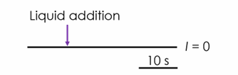
Protocol B – current clamp protocol for QPatch 48
Zero current was used to record the cell resting membrane potential (RMP) for 40 s per liquid period (Protocol B). Liquid addition occurred after 10 s recording and the effect on RMP was assessed 30 s thereafter. Vehicle (≤ 1 % DMSO) was applied, followed by four cumulative concentrations of compound. Recordings were conducted using single hole acquisition.
Manual patch clamp recordings from sensory neurons
Neonatal Wistar rat neurons (P4-P12) were isolated, dissociated and plated at appropriate densities onto poly-D-lysine- and laminin-coated glass coverslips. Manual patch clamp experiments were performed using a MultiClamp 700B amplifier (Molecular Devices) and an Axon Digidata 1400A digitiser (Molecular Devices).
1. Potency assessment of Kv7.2/7.3 activators using a Fluorescent Imaging Plate Reader (FLIPR)
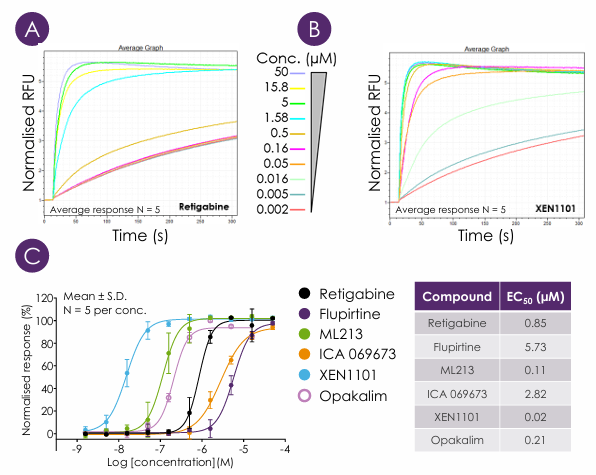
Figure 1 – Potency of KV7.2/7.3 activators assessed using a FLIPR Penta thallium flux assay. Averaged (N = 5) raw data traces for retigabine (A) and XEN1101 (B). The legend for (B) is the same as for (A). RFU = relative fluorescence units. (C) EC50 concentration-response curves normalised to 0.5 % DMSO and 50 µM retigabine.
2. Electrophysiological characterisation of Kv7.2/7.3 activator modalities using Qube384
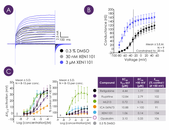
Figure 2 – Pharmacological effects of KV7.2/7.3 activators characterised using automated voltage clamp electrophysiology. (A) Example I-V current traces of 0.3 % DMSO and 3 µM XEN1101. (B) Average conductance voltage curves for 0.3 % DMSO (V0.5 = -27.70 mV – dotted line), 30 nM (V0.5=-28.40mV) and 3 µM (V0.5=-65.15mV) XEN1101. The legend for (B) is the same as for (A). (C) EC50 potency assessment of compounds on KV7.2/7.3 V0.5 activation and current (I) at +50 mV.
3. Physiological effect of KV7.2/7.3 activators on cell resting membrane potential (RMP) using QPatch 48
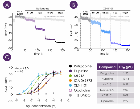
Figure 3 – KV7.2/7.3 activators hyperpolarise the RMP of U2-OS-KV7.2/7.3 cells. Raw current clamp traces from retigabine (A) and XEN1101 (B). Each panel also shows a time-matched vehicle control trace (1 % DMSO). (C) EC50 potency assessment of KV7.2/7.3 activators on RMP. Maximal hyperpolarisation approaches EK.
4. Effects of KV7.2/7.3 activators on sensory neuron resting membrane potential (RMP)
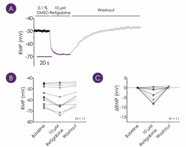
Figure 4 – Sensory neuron hyperpolarisation by retigabine. (A) Example trace showing sensory neuron RMP hyperpolarisation in response to 10 µM retigabine. Graphs illustrate raw RMP (B) and ∆RMP (C) in response to baseline (0.1 % DMSO), 10 µM retigabine and washout. Cell-to-cell variability in retigabine response was observed.
A comprehensive understanding of the pharmacology of KV7.2/7.3 activators provides a route to developing more potent and efficacious KV7.2/7.3-selective compounds for the treatment of pain and epilepsy.
We have developed an integrated workflow combining high-throughput screening using a FLIPR thallium dye assay, Qube 384 voltage clamp profiling to characterise biophysical effects of KV7.2/7.3 activators, followed by QPatch 48 current clamp studies to assess physiological effects on resting membrane potential. Key compounds can then be triaged for validation in sensory neuron recordings, linking pharmacological mechanism to neuronal excitability.
Action Potential Waveform Analysis in Human IPSC-Cardiomyocytes Enables Mechanistic Assessment of Multichannel Cardiac Effects
Cardiac toxicity remains the leading cause of new drug safety side-effects. Current preclinical cardiac safety assays rely on in vitro cell-based ion channel assays and ex vivo and in vivo animal models⁽¹⁾. These assays provide an indication of acute risk but they do not always predict the effect of chronic compound exposure, as recently seen with oncology drugs. Therefore, new assays are required to characterise chronic structural and functional effects in human cells earlier in drug discovery. Impedance-based technology can provide more accurate chronic cardiotoxicity measurements in an efficient manner using human induced pluripotent stem cell-derived cardiomyocytes (hiPSC-CMs).