
Define clinical exposure associated with QTc liability

Data Card

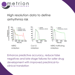
High resolution data to define arrhythmia riskEnhance predictive accuracy, reduce false negatives and late-stage failures for safer drug development with improved preclinical-to-clinical translation

Recordings using voltage-sensitive dye (VSD) with quality equivalent to patch-clamp are crucial for assessing cardiovascular risk in drug discovery. They provide a high-fidelity, high-throughput alternative to traditional electrophysiology techniques.

VSD-based recordings offer non-invasive, high-resolution measurements of membrane potential changes across a large number of cells or wells simultaneously, enabling faster and more efficient cardiac safety screening. This approach ensures precise detection of drug-induced effects on cardiac ion channels, such as hERG (IKr), Nav1.5, and Cav1.2, which are critical for evaluating proarrhythmic risk.

The ability to achieve patch-clamp-equivalent data quality using VSD enhances predictive accuracy, reducing false negatives and late-stage failures, and supports safer drug development with improved preclinical-to-clinical translation.

Read more about our hiPSC cardiomyocyte assay.

Contact us for a quote or discussion


Recommended Publications
Latest Publications
An Integrated Assay Platform for Mechanistic and Translational Profiling of Kv7.2/7.3 Activators

An integrated assay platform was developed to characterise the pharmacological and physiological effects of Kv7.2/7.3 activators. Combining high-throughput FLIPR thallium flux, automated voltage- and current-clamp electrophysiology, and sensory neuron recordings enabled assessment of compound potency, mechanism of action, and effects on neuronal resting membrane potential. Profiling of retigabine, flupirtine, ML213, ICA 069673, XEN1101 and opakalim demonstrated distinct pharmacological profiles across complementary assay systems. The workflow provides a mechanistic and translational approach for characterising Kv7.2/7.3 activators and their effects on neuronal excitability.

Action Potential Waveform Analysis in Human iPSC-Cardiomyocytes Enables Mechanistic Assessment of Multichannel Cardiac Effects

Optical voltage imaging of human iPSC-derived cardiomyocytes was used to assess electrophysiological effects of compounds beyond hERG inhibition. Action potential waveform analysis revealed compound-specific and concentration-dependent changes, enabling mechanistic differentiation of multichannel activity and demonstrating a human-relevant approach for translational cardiac safety assessment.

View All
magnifier
linkedin facebook pinterest youtube rss twitter instagram facebook-blank rss-blank linkedin-blank pinterest youtube twitter instagram