By Federico Olivero, Research Associate Scientist, Metrion Biosciences
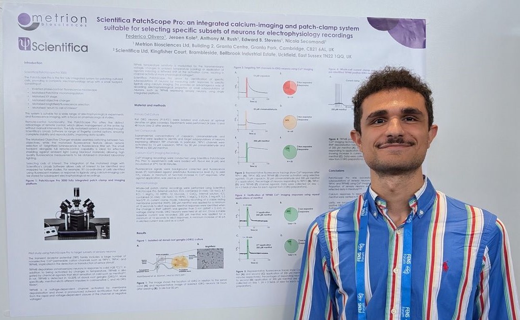
It was a pleasure to present this poster at the FENS Forum. The poster details Metrion and Scientifica’s1 collaboration on the optimisation and utilisation of the PatchScope Pro2. We first targeted three different TRP channels (TRPV1, TRPA1 and TRPM8) in rat dorsal root ganglia (DRGs). Subsequently we focused on TRPM8 which is a multimodal sensor (temperature, pressure, voltage, osmolality) present in 10-20% of DRGs neurons.
Using calcium-imaging, response of DRG neurons to TRPV1, TRPA1 and TRPM8 ligands were tested, where 64% of neurons responded to 10 µM capsaicin, 29% to 30 µM cinnamaldehyde and 13% to 500 µM menthol. Subsequently a compound-application protocol was optimized using repeat applications of 200 µM menthol, suitable for combined calcium-imaging/electrophysiology. TRPM8 expressing neurons were characterized using electrophysiology following initial selection by calcium-imaging (menthol positive), where effects of 200 µM menthol were demonstrated in both voltage-clamp and current-clamp recordings (n=5). This study demonstrates the advantages of the PatchScope system for selecting specific subsets of neurons using calcium-imaging prior to electrophysiology recordings.
- https://www.scientifica.uk.com.
- Scientifica’s PatchScope Pro is an integrated electrophysiology rig incorporating an inverted phase-contrast fluorescence microscope, motorised XY stage and PatchStar micromanipulators, suitable for patch-clamp recording. In contrast to other patch-clamp systems, this microscope has a small footprint to maximise use of lab space. The low-noise electrical components are designed specifically for patch-clamp recordings. Fluorescence capability allows identification of cells of interest (e.g. using fluorescent markers or calcium-imaging) and motorisation of the stage means that XY co-ordinates of cells can be stored for subsequent electrophysiology and imaging.

